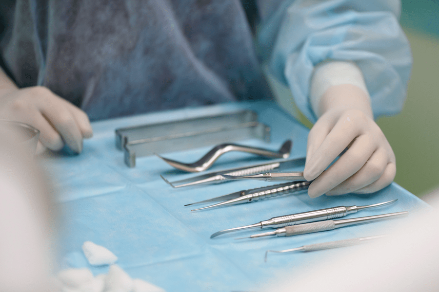
Dental Surgery
Accommodation

Anasthesia

Length of procedure

Companionship

Bone Graft
A dental bone graft adds volume and density to your jaw in areas where bone loss has occurred. The bone graft material may be taken from your own body (autogenous), or it may be purchased from a human tissue bank (allograft) or an animal tissue bank (xenograft). In some instances, the bone graft material may be synthetic (alloplast).
Once the bone graft has been placed, it holds space for your own body to do the repair work. In other words, a dental bone graft is like a scaffold on which your own bone tissue can grow and regenerate. In some cases, your dental provider may combine a dental bone graft with platelet-rich plasma (PRP). This is taken from a sample of your own blood and is used to promote healing and tissue regeneration.
A person with bone loss in their jaw usually needs a dental bone graft. This procedure may be recommended if you:
Are having a tooth extracted.
Plan to replace a missing tooth with a dental implant.
Need to rebuild the jaw before getting dentures.
Have areas of bone loss due to gum (periodontal) disease.
First, your dental provider will numb the area with local anesthetic. Next, they'll create a small incision in your gums. Gum tissue is moved back slightly so that the jawbone is visible. After cleaning and disinfecting the area, your dentist adds bone grafting material to repair the defect. In many cases, the bone graft is covered with a membrane for additional protection. Finally, the gum tissue is repositioned and the incision is closed with stitches.
Following a dental bone graft, you may have pain, swelling and bruising. These are normal side effects that should diminish in a few days. Symptoms can be managed with pain relievers. Your dentist may give you antibiotics as well. These should be taken exactly as prescribed. You might notice small fragments of bone coming out of the site over the first few days. These pieces often resemble grains of salt or sand. This usually isn’t a cause for concern, but call your dentist to make sure that you’re healing as expected.
Most people who have dental bone grafts report little to no pain. Just be sure you take all medications as prescribed and follow your post-operative instructions closely.
As mentioned above, recovery times can vary significantly for each person. Once the bone graft is placed, Viva La Dent Clinic dentist’s will monitor your healing by getting X-ray. If you’re waiting to undergo dental implant surgery, they will let you know when your new bone is strong enough to support the implant.
Dental Bone Augmentation
Dental bone augmentation is a valuable technique in modern dentistry, enabling patients with inadequate bone structure to benefit from dental implants and other restorative dental procedures, which can significantly enhance their oral health and quality of life. However, the suitability of the procedure and the choice of graft material should be carefully evaluated by a dental professional based on the individual's specific needs and circumstances. Dental bone augmentation, also known as bone grafting in dentistry, is a surgical procedure used to increase the amount or quality of bone in the jaw or oral cavity. This procedure is typically performed when a patient lacks sufficient bone density or volume to support dental implants or other dental prosthetics. Dental bone augmentation can help create a stable foundation for dental implants, improve the aesthetics and functionality of the teeth, and restore the oral health of a patient. Here are some key aspects of dental bone augmentation:
Dental Implants
One of the most common reasons for dental bone augmentation is to prepare the jawbone to receive dental implants. A solid and stable jawbone is essential for the long-term success of dental implant placement.
Tooth Extractions
After a tooth extraction, the surrounding bone may undergo resorption or shrinkage. Augmentation can help preserve the bone for future implant placementor denture support.
Trauma or Disease
Dental bone augmentation may be necessary after traumatic injuries, infections, or diseases that have led to bone loss in the oral cavity.
Types of Bone Grafts
Several types of bone graft materials can be used in dental bone augmentation, including: Autografts: Bone taken from the patient's own body, often from another part of the jaw, hip, or tibia. Allografts: Donor bone tissue from another human source, typically from a tissue bank. Xenografts: Bone tissue sourced from non-human animals, usually bovine or porcine.
Synthetic Grafts
Man-made materials designed to stimulate bone growth, such as calcium-based ceramics.
Procedure
The procedure involves making an incision in the gum tissue to access the deficient bone area. The graft material is then placed in the target area, and the gum tissue is sutured closed. Over time, the patient's natural bone should integrate with the graft material, creating a stronger and denser bone structure.
Healing and Recovery
Healing time varies depending on the individual and the extent of the grafting procedure. It may take several months for the graft to fully integrate with the existing bone. During this healing period, patients may need to follow a soft diet and avoid putting excessive pressure on the treated area.
Success and Follow-Up
The success of dental bone augmentation is determined by the integration of the graft material and the ability of the new bone to support dental prosthetics. Follow-up appointments with the Viva La dentist’s or oral surgeon are essential to monitor the healing process and ensure the success of the procedure.
Connective Tissue Graft
A dental connective tissue graft, also known as a subepithelial connective tissue graft or simply a connective tissue graft, is a surgical procedure used in dentistry to treat gum recession and improve the aesthetics and health of the gums. This procedure involves taking a small piece of connective tissue, often from the roof of the mouth (palate), and transplanting it to the area where gum recession has occurred. Here's an overview of dental connective tissue grafting: Fortunately, several treatments for gum recession are available, depending on the severity of tissue loss. Early detection is key, as earlier diagnosis and treatment lead to better outcomes. Gum recession may be caused by several different factors, including but not limited to:
Aggressive brushing over the long term
Diabetes
Family history of gum disease
HIV
Hormonal Changes in women
Smoking
Tartar
Certain medications may also cause dry mouth, which increases one's risk of gum recession. Gum recession is most common in adults aged 40 and older.
Apical Resection Surgery
Apical Surgery is a minor surgical procedure carried out to treat a tooth which has persistent infection. This may arise when root canal treatment has not been completely successful. The infection is often found around the tip of the root of the tooth, within the jaw bone. It is usually carried out under local anaesthesia (i.e. after being numbed up) with the patient awake. You can go home immediately after the procedure and many patients do not need to take any time off work after the procedure.
Infection at the tip of the root (also known as the root apex) usually arises from within an infected root canal (the thin canal within the root, where the nerve and blood vessels are). Usually root canal treatment (a root filling) is sufficient to eliminate this infection but apical surgery may be necessary in the following situations:
When root canal treatment has been unsuccessful and repeating the root filling is not possible or may not work.
When the root filling has been repeated but has been unsuccessful.
When there are bits of root filling material, which have been pushed through the root apex and cannot be retrieved.
When a biopsy of the infected area is required.
An incision is made around the tooth and the gum is gently peeled back. The dentist would then normally be able to see the part of the jaw bone where the infection is situated. All infected tissue would then be removed.
Using a drill, the tip of the root will then be carefully removed and a special filling would then be placed over the freshly cut root to seal it. Placing this filling reduces the risk of the infection returning.
The gum is then stitched back into place.
Apical surgery is often carried out when other options to save the tooth have been exhausted. Therefore, the success rate varies significantly on the condition of the tooth and previous treatment(s) that have been carried out. On average, the success rate can be between 75% and 85%.
Get a free offer now!
We tell you the total cost of your treatment from the beginning and then avoid any additional costs. You can be assured that there will be no extra cost, except the total cost of dental treatment sent to you. You will be answered by both email & WhatsApp.
Sinus Lifting (Sinus Augmentation)
Sinus lifting, also known as a sinus augmentation or sinus elevation, is a surgical procedure performed in dentistry to increase the amount of bone in the upper jaw's posterior (back) region, particularly in the area of the maxillary sinuses. The maxillary sinuses are air-filled spaces located above the upper molars and premolars. Sinus lifting is typically done to create a more stable foundation for dental implants in cases where there is insufficient natural bone in this region. Here's an overview of sinus lifting: In order for a dental implant to be successful, the quantity and quality of the bone where the implant would be placed must be sufficient. It is not uncommon for patients to lose bone in the upper jaw, specifically between the molars and premolars — which is also the same space between the jaw and maxillary sinuses on either side of the nose. This bone loss may be due to a maxillary sinus too close to the upper jaw, reabsorption, periodontal disease, or tooth loss. Consequently, patients may have an insufficient quality bone for implant placement. In sinus lift surgery, also known as sinus augmentation, a periodontist lifts the sinus floor and develops new bone for dental implant placement. It is a complex procedure that should only be performed by a dental specialist. Sinus lift surgery is an especially appealing option for those whose only tooth replacement options would be loose dentures. Loose dentures are less advantageous than dental implants, as they do not particularly look or function like natural teeth. Dental implants are also better for jawbone health in the long run.
Patients should place a gauze pad over the surgical area for a half-hour immediately after surgery. How successful are sinus lifts? Sinus lifts have an overall high success rate. One study found that, after 24 months of evaluation, dental implants in sinus lifts reached a success rate of 95.2%. This success rate was high as 100% for those with a residual bone height of 4 mm or higher and high as 87.5% for those with a residual bone height under 4 mm.
While sinus lifts are a generally safe procedure, every surgery comes with a certain set of risks. The main concern with a sinus lift is the possibility of puncturing or tearing the sinus membrane. However, if this does occur, the periodontist should be able to remedy it by stitching the sinus tear or patching it. If this is insufficient, the periodontist may need to stop the surgery to allow the hole to heal. Infection is another, yet uncommon, risk. Infection and rejection of the bone graft are also rare risks intrinsic to the procedure.